
Buildings at the John A. Burns School of Medicine campus. The CCR base is located on the third floor of the Biosciences Building.
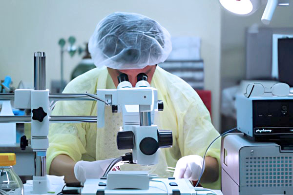 At the Kakaʻako campus we have been able to develop important core facilities shared with all of the approximately 100 investigators working here. NIH funding from the Diabetes COBRE, as well as institutional support from the medical school and the offices of the Vice-Chancellor and Vice President for Research, support a Mouse Phenotyping Core.
Equipment is available upon contacting a CCR Investigator for training and availability.
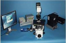 The IonOptix imaging system offers a fast real-time, turnkey system for recording myocyte calcium and contractility, with simultaneous acquisition of fluorescence photometry and digital cell geometry measurements.
Contact: Dr. Matsui
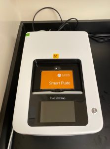 The Maestro Pro multiwell microelectrode array (MEA) system allows for non-invasive evaluation of cells in an easy-to-use in vitro assay. Whether monitoring the intricate, electrical activity of excitable cells (e.g. neurons and cardiomyocytes), or tracking the growth and death of cancer cells with optional addon modules, Maestro Pro allows researchers to investigate the functionality of cells in a multiwell plate.
Contact: Dr. Zhang
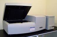 Typhoon™ 9410 Variable Mode Imager unites proven storage phosphor autoradiography technology with four-color, non-radioactive fluorescent labeling techniques. For DNA, RNA, and protein samples, choose from: storage phosphor autoradiography, direct blue-excited fluorescence (457, 488 nm), direct green-excited fluorescence (532 nm), direct red-excited fluorescence (633 nm), and chemiluminescence.
Contact: Dr. Shohet
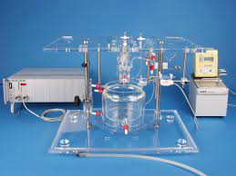 Langendorff: The CCR has two independent Langendorff systems for ex vivo heart study and cardiomyocyte isolation.
Contact: Dr. Matsui or Dr. Shohet
Beckman Avanti-JE floor centrifuge with 500ml and 50ml fixed angle rotors, as well as a swinging bucket rotor with various adapters.
Contact: Dr. Shohet
AKTA prime FPLC
Contact: Dr. Boisvert
Gel Doc EZ Imager Automated Gel Imaging Instrument (BIO-RAD )
Atomic Force Microscope
The CCR coordinates with a number of external facilities that provide core or specialized services in other UH departments.
The Genomics and Bioinformatics Shared Resource (GBSR) at JABSOM and UH Cancer Center (UHCC).
https://www.uhcancercenter.org/research/shared-resources/genomics-and-bioinformatics
The Flow Cytometry Facilities equipped with state-of-the-art equipment for cellula analysis and sorting is operated by the Shared resources at the JABSOM and the UH Cancer Center. Also available is the equipment and services from the School for Ocean, Earth Sciences and Technology Flow Cytometry Facility.
Department of Quantitative Health Science (QHS) provides a range of customized statistical and database management services and training for biomedical researchers and clinicians at the John A. Burns School of Medicine.
https://qhs.jabsom.hawaii.edu/
The Biological Electron Microscope Facility (BEMF) at the University of Hawaiʻi is a multi-user/service facility, administered by the Pacific Biosciences Research Center (PBRC).
http://bemf.pbrc.hawaii.edu
Koa, the High Performance Computing (HPC) cluster at the University of Hawai‘i (UH), is a collection of many computers nodes connected together with a network that solves computational problems which are too large for standard computers. UH Information Technology Service Cyberinfrastructure (CI) operates Koa as a free UH system-wide computational resource that supports data and computationally intensive research in over 90 disciplines.
https://datascience.hawaii.edu/hpc/
Acoustic microscopy
https://www.soest.hawaii.edu/~zinin/Zi-SAM.html
In addition to the CCR’s Semiconducter Paramertic Analyzer (SPA) capabilities, SPA testing is available through the School of Engineering
Contact: Dr. Aaron Ohta
Chemistry Department Services